Labeled Anterior And Posterior Muscles Of The Body / Muscles Of The Trunk Anatomy Diagram Pictures Kenhub / Behind the rhomboids are on the posterior aspect of the body.
byBen Potts-
0
Labeled Anterior And Posterior Muscles Of The Body / Muscles Of The Trunk Anatomy Diagram Pictures Kenhub / Behind the rhomboids are on the posterior aspect of the body.. Our muscles of the leg quizzes and labeled diagrams might be. It is locatedlocated directly anterior to a groove between the femur condyles called the patellar surface. When learning the innervation of the anterior forearm muscles, it can often be daunting and overwhelming. Behind the rhomboids are on the posterior aspect of the body. There are approximately 680 skeletal muscles within the typical human, and almost every muscle constitutes one part of a pair of identical bilateral muscles, found on both sides, resulting in approximately 320 pairs of muscles, as presented in this article.
Knowing which muscles are in the anterior of the body vs posterior is key to answering several questions in both the level 2 and level 3 anatomy and some muscle names indicate the number of muscles in a group. The tibialis posterior muscle is one of the small muscles of the deep posterior compartment of the leg. Nevertheless, the exact number is. Upper half of posterior shaft of superficial slip inserts on the tuberosity of the navicular bone and sometimes medial cuneiform. Anterior and posterior pulmonary plexus of nerves.
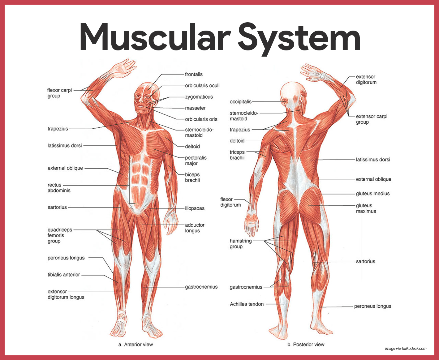
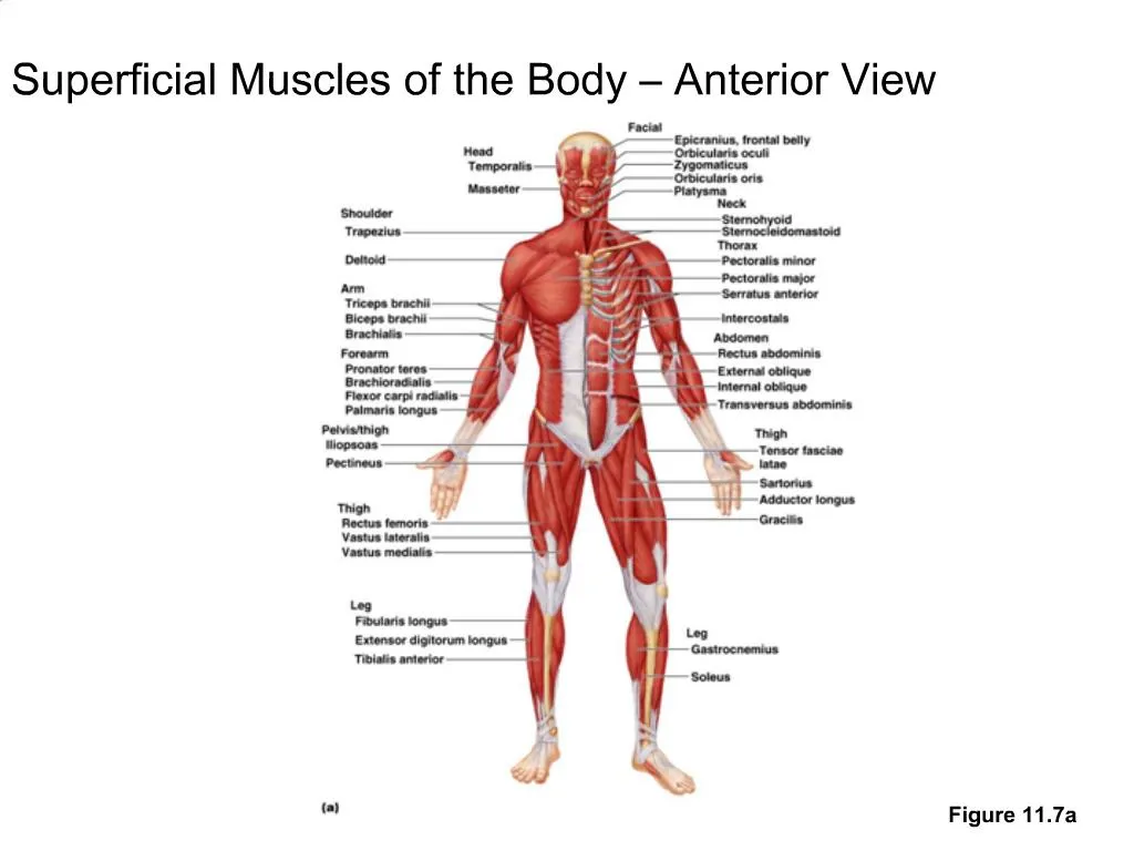
Muscular System Anatomy And Physiology Nurseslabs from nurseslabs.com The muscles labelled in the anterior muscles diagram shown above are listed in bold in the following table The labeled structures (listed alphabetically) are: Muscles of the ankle and foot. Most of the important bones and groups of bones in the human body are visible in the anterior view of the. Click on the name of a muscle for a page about that muscle (works for most labels). There are around 650 skeletal muscles within the typical human body. Superficial fascia of anterior abdominal wall: Superficial and deep posterior muscles of upper body.
The skeleton is an aggregate of many connected bones.
Anterior muscles of lower leg. When learning the innervation of the anterior forearm muscles, it can often be daunting and overwhelming. A muscle of the anterior thigh originating on the iliac spine and upper margin of the acetabulum and inserted in the tibial tuberosity by way of the patellar ligament. Bones are hard but alive those that cannot be seen lie within the skull. The tibialis anterior, extensor this muscle is the most posterior and lateral of all the muscles of the anterior leg. Two facets (or depressions) on the posterior side of the patella articulate the patella is embedded in the quadriceps tendon, which makes it the largest sesamoid (= tendon embedded) bone in the body. The 2 recti draw the head backward. At the level of whitnall's ligament the palpebral segment of the levator palpebrae superioris splits into an anterior and posterior division Limb in posterior human body. The anterior and posterior axillary lines drop vertically from the anterior and posterior axillary folds (the muscles that border the axilla). Our muscles of the leg quizzes and labeled diagrams might be. It is the most superficial of the calf muscles. Divides the body or any of its parts into anterior and posterior portions.
Foreign body, lung tumor, lobar pneumonia, pleural effusion, pneumothorax, partial paralysis of the respiratory muscles. At the level of whitnall's ligament the palpebral segment of the levator palpebrae superioris splits into an anterior and posterior division The labeled structures (listed alphabetically) are: Do you prefer a more interactive learning approach? It is the most superficial of the calf muscles.
Ppt Superficial Muscles Of The Body Anterior View Powerpoint Presentation Id 526126 from image4.slideserve.com Which organ is responsible for pumping blood around the body? Toward the head or upper part of a structure: Behind the rhomboids are on the posterior aspect of the body. Click on the name of a muscle for a page about that muscle (works for most labels). Towards or on the back of the body: Putting this in context, the heart is posterior to the sternum because it lies behind it. The deep muscles of the back and the suboccipital muscles are supplied by the posterior primary rami of the spinal nerves. Muscle around mouth that allows smiling muscular system with posterior and anterior skeletal muscles labeled.
The tibialis anterior, extensor this muscle is the most posterior and lateral of all the muscles of the anterior leg.
The 2 recti draw the head backward. Most of the important bones and groups of bones in the human body are visible in the anterior view of the. The bones of the skeletal system act as attachment points for the skeletal muscles of the body. The labeled structures (listed alphabetically) are: You will also find anus, metanephridium, intestine, gizzard, ventral nerve cords with segmental ganglia, circulatory system, subpharyngeal ganglion, the mouth of earthworm, cerebral ganglia, pharynx, esophagus, clitellum, crop part in anatomical regions of the human body. Support and protect the abdominal viscera. Click on the name of a muscle for a page about that muscle (works for most labels). The serratus anterior is below the axilla, on the lateral part of the it originates on the upper eight or nine ribs on the lateral and anterior thorax and inserts in the scapula on the side toward the vertebrae. The tibialis anterior, extensor this muscle is the most posterior and lateral of all the muscles of the anterior leg. The deep muscles of the back and the suboccipital muscles are supplied by the posterior primary rami of the spinal nerves. • he allowed his beloved cousin patroclus to fight in his armor, and when hector slew patroclus, achilles returned to battle, killed hector, and dragged his body around the walls of troy. Which organ is responsible for pumping blood around the body? • muscles of the body can be broadly classified based on structure, contractile properties, control mechanisms into.
The labeled structures (listed alphabetically) are: The 2 recti draw the head backward. Which organ is responsible for pumping blood around the body? Anterior refers to the 'front', and posterior refers to the 'back'. Most of the important bones and groups of bones in the human body are visible in the anterior view of the.
Https Www Pearsonhighered Com Assets Samplechapter 0 1 3 4 013439495x Pdf from The muscles found in the anterior compartment of the leg are: This muscle diagram is interactive: Toward the head or upper part of a structure: Arises from medial aspect of anterior 2/3rd of the zygomatic arch & from the lower border of the posterior 1/3rd of the zygomatic arch. Associated structures are labeled in parentheses. An example of this is the quadriceps, a group of four muscles located on the. .muscle diagram of the back posterior front anterior : The anterior and posterior axillary lines drop vertically from the anterior and posterior axillary folds (the muscles that border the axilla).
Divides the body or any of its parts into anterior and posterior portions.
Muscle of the forehead that moves the forehead skin and eyebro… ring muscle of the eye socket; Muscles transfer force to bones through tendons. Anterior muscles of lower leg. The deep muscles of the back and the suboccipital muscles are supplied by the posterior primary rami of the spinal nerves. Above the humerus is superior to the radius. Bo., bowman's layer the longitudinal muscle of the ciliary body attaches to the scleral spur and opens the trabecular it is the anterior border of the trabecular meshwork and the posterior border of descemet's membrane. Nevertheless, the exact number is. The 2 recti draw the head backward. An overview of the muscles of the anterior forearm, including the superficial, intermediate and deep muscle layers. The longus colli muscle is situated on the anterior surface of the vertebral column, between the atlas and the third thoracic vertebra. Arises from medial aspect of anterior 2/3rd of the zygomatic arch & from the lower border of the posterior 1/3rd of the zygomatic arch. At the level of whitnall's ligament the palpebral segment of the levator palpebrae superioris splits into an anterior and posterior division The muscles labelled in the anterior muscles diagram shown above are listed in bold in the following table
Limb in posterior human body anterior muscles of the body labeled. Posterior to the orbicularis muscle lies the orbital septum, with the conjunctival epithelium forming the posterior aspect of the eyelid.